How do we breathe? (Lungs and Pleura) Interactive Biology, with Leslie Samuel
Total Lung Capacity (TLC) and Lung Compliance. TLC refers to the maximum volume of air the lungs of an adult person can hold. It is the sum of the air released by the lung after a maximum exhalation (vital capacity or VC) and the volume of air left behind within the lungs after a deepest exhalation (residual volume or RV) [46].The TLC of human lungs is 6 liters [47].
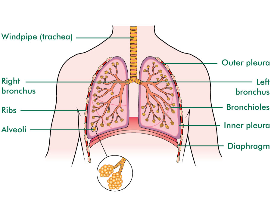
The lungs Macmillan Cancer Support
Lower Genitourinary Trauma. The purpose of the lung is to provide oxygen to the blood. The respiratory system divides into airways and lung parenchyma. The airways consist of the bronchus, which bifurcates off the trachea and divides into bronchioles and then further into alveoli.

Foods to help keep your lungs and respiratory system healthy
The apex is the tip of the nose. On either side of the apex, the nostrils are formed by the alae (singular = ala). An ala is a cartilaginous structure that forms the lateral side of each naris (plural = nares), or nostril opening. The philtrum is the concave surface that connects the apex of the nose to the upper lip.
Respiratory system Canadian Lung Association
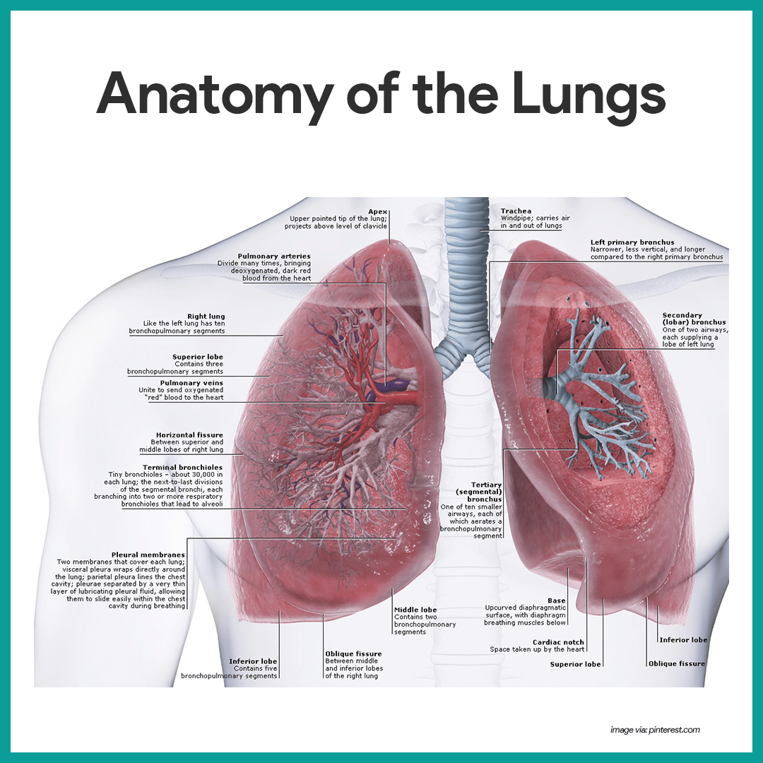
Lung Structure The lungs are roughly cone shaped, with an apex, base, three surfaces and three borders. The left lung is slightly smaller than the right - this is due to the presence of the heart. Each lung consists of: Apex - The blunt superior end of the lung. It projects upwards, above the level of the 1st rib and into the floor of the neck.

Lung Structure BioNinja
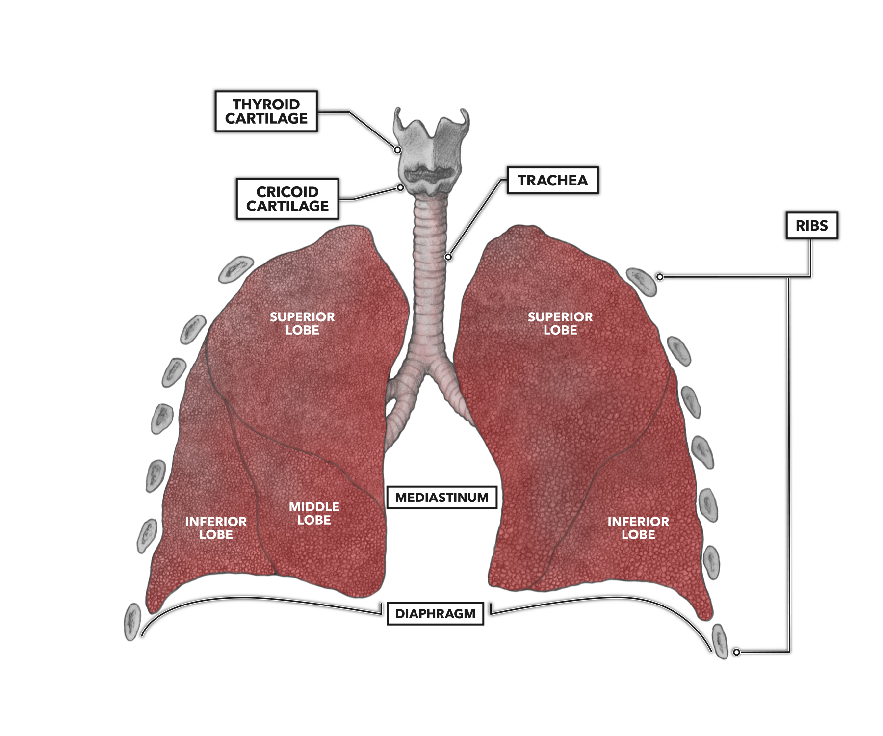
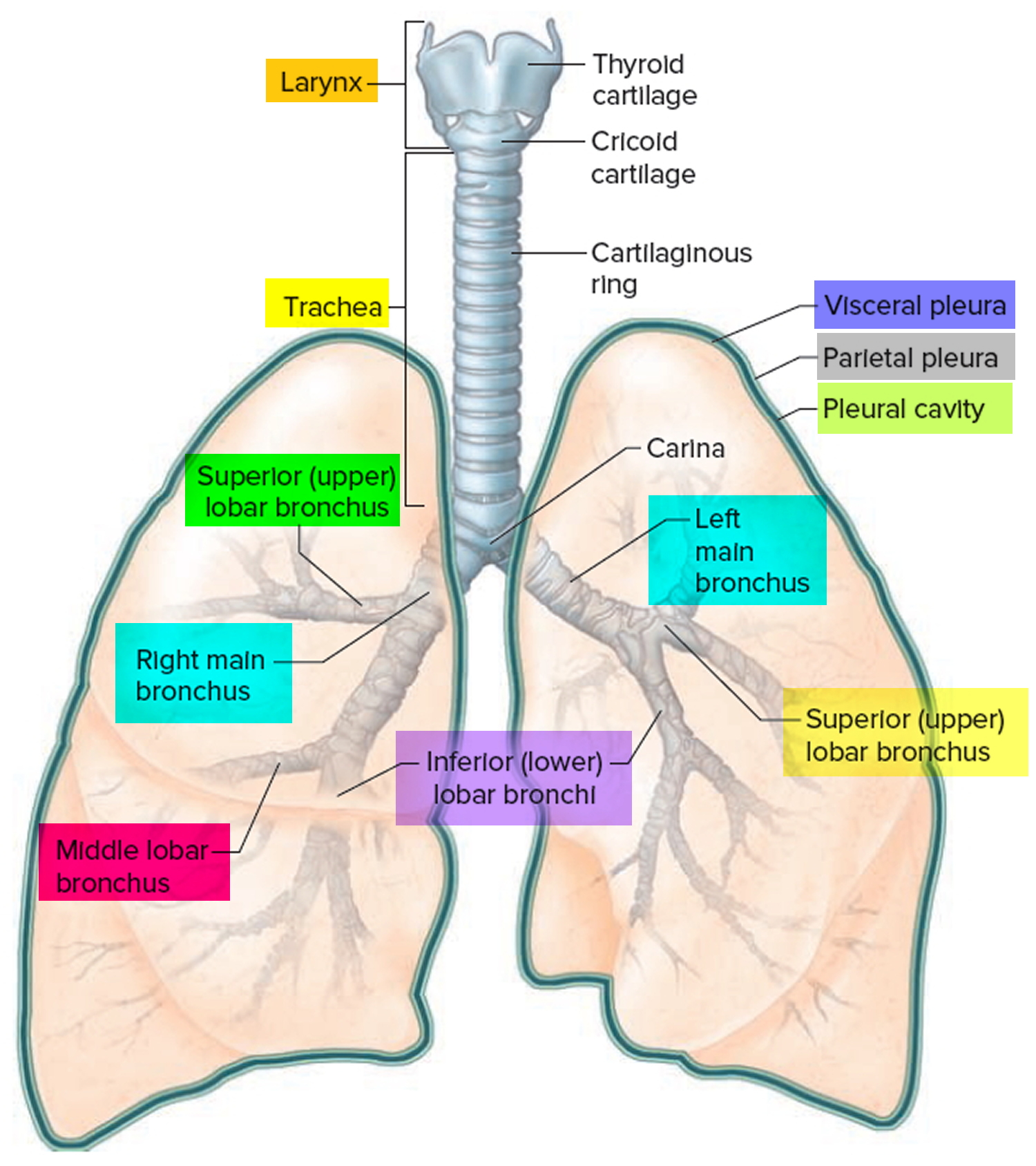
Gross Anatomy of the Lungs. The lungs are pyramid-shaped, paired organs that are connected to the trachea by the right and left bronchi; on the inferior surface, the lungs are bordered by the diaphragm. The diaphragm is the flat, dome-shaped muscle located at the base of the lungs and thoracic cavity.
The lungs Lung cancer Macmillan Cancer Support
Functionally, the lung is divided into a series of bronchopulmonary segments. The bronchopulmonary segments are the largest subdivision of a lobe. They are separated from adjacent segments by connective tissue septa and are also surgically resectable. They are 10 bronchopulmonary segments in the left lung and 8-10 in the left lung [9].
The structure of a lung with labeled parts. Biology vector illustration vector de Stock Adobe
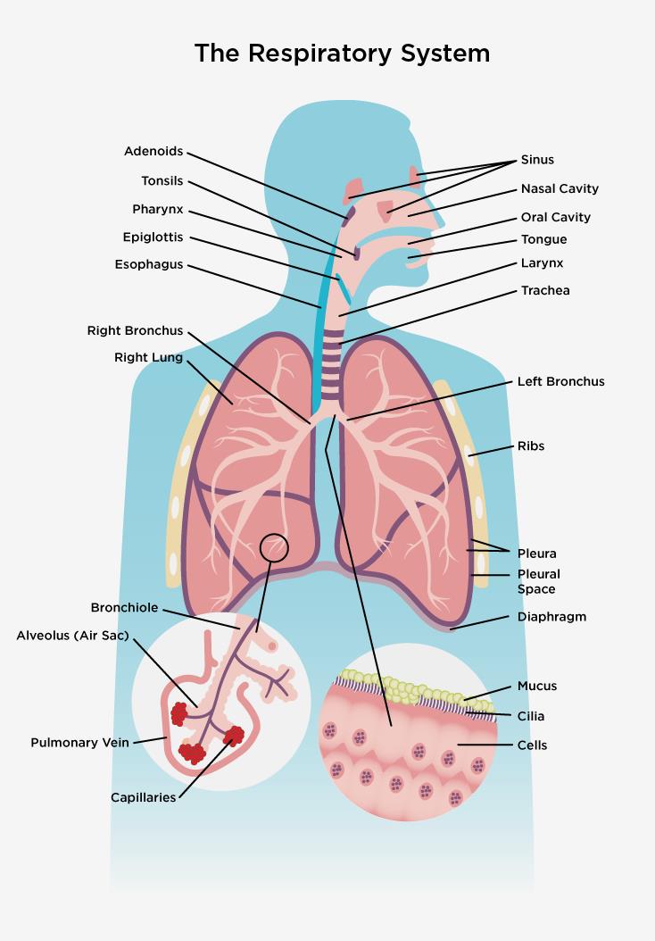
The respiratory system, also called the pulmonary system, consists of several organs that function as a whole to oxygenate the body through the process of respiration (breathing).
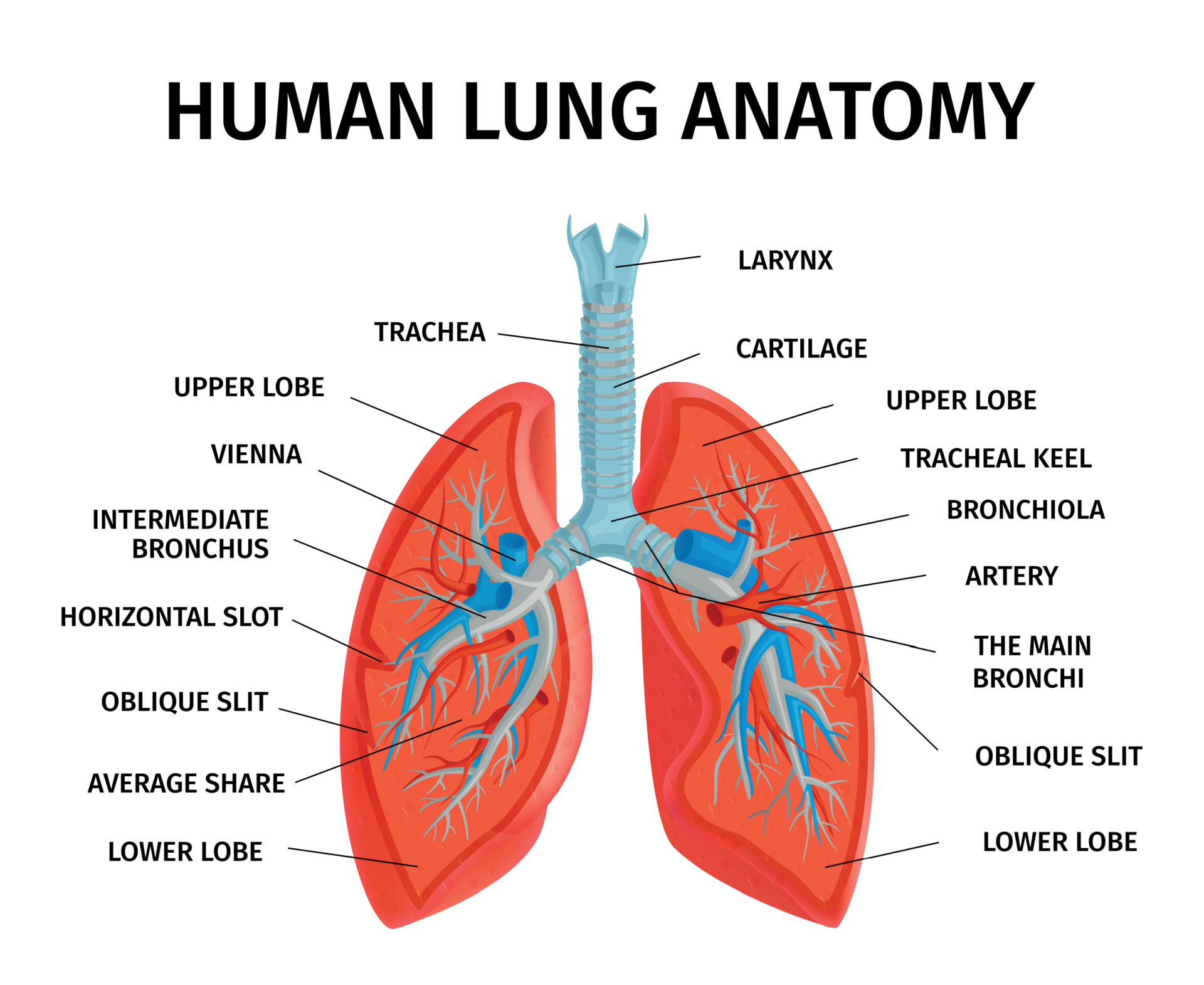
Human Lung Anatomy Diagram 4958464 Vector Art at Vecteezy
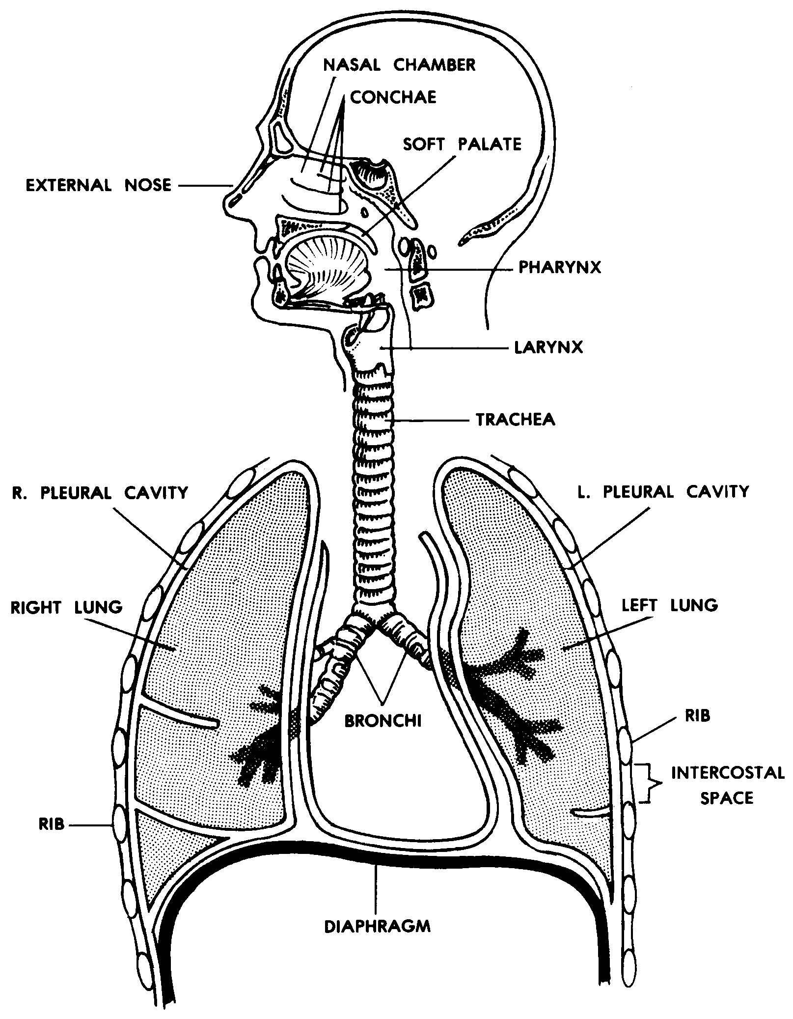
The human lungs are a pair of spongy organs within the thoracic cavity that facilitate gaseous exchange. They are a part of the respiratory system, which also includes the nose, nasal sinuses, mouth, pharynx, larynx, and trachea. At the level of the lungs, much-needed oxygen is absorbed into the blood, while waste gases are excreted and exhaled.

Sections of the Lungs Seattle Cancer Care Alliance
Lungs Diagram in Human Body Humans have a right and a left lung positioned in the chest cavity. Jointly, the lungs inhabit most of the intrathoracic space. Lungs are responsible for adding oxygen and removing carbon dioxide from the blood, thus serving as a gas-exchanging structure for respiration.
A healthy lung has a pinkish appearance, and if you could see it outside the body, it would look
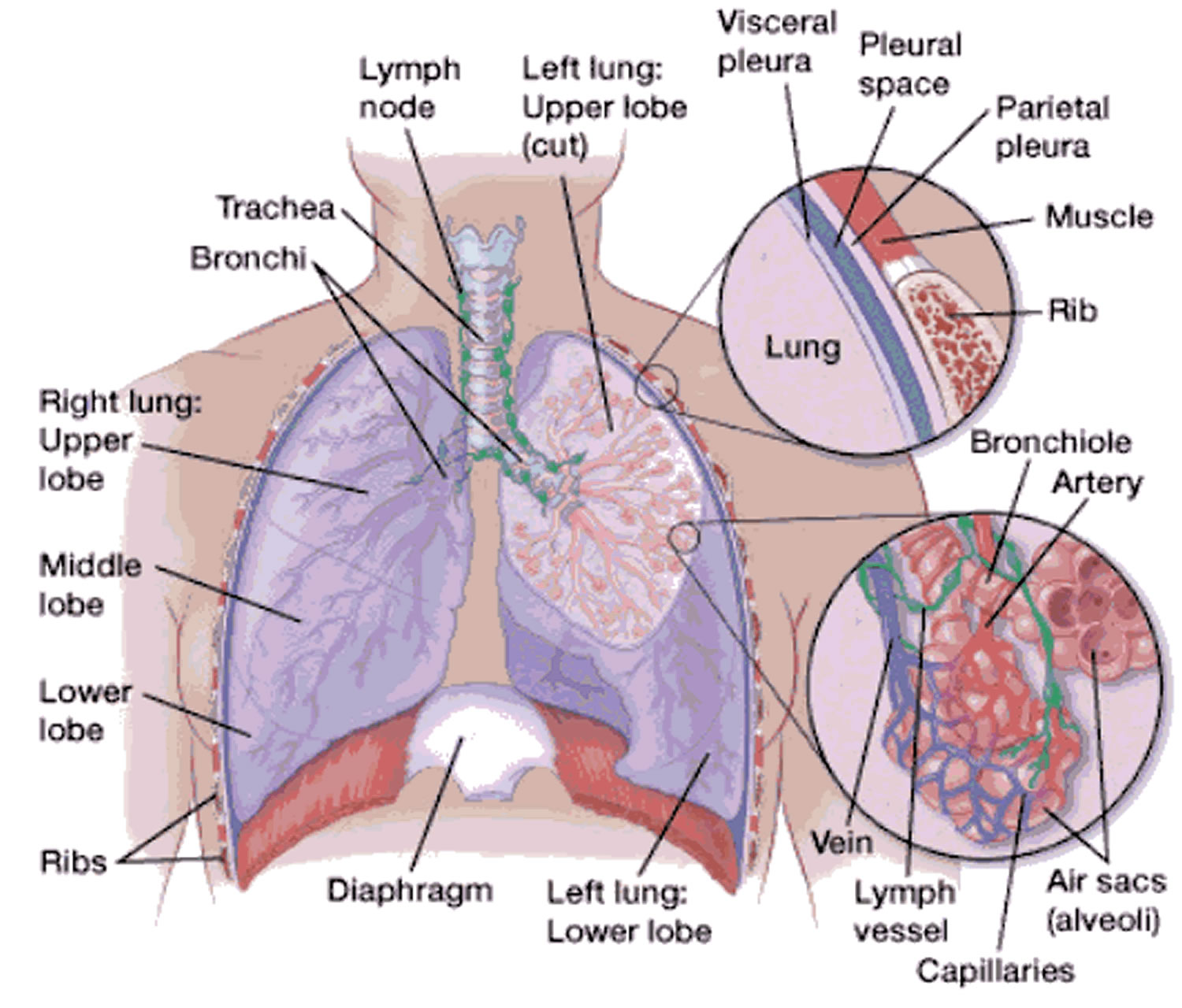
The diaphragm is the flat, dome-shaped muscle located at the base of the lungs and thoracic cavity. The lungs are enclosed by the pleurae, which are attached to the mediastinum. The right lung is shorter and wider than the left lung, and the left lung occupies a smaller volume than the right.

Respiratory System Anatomy
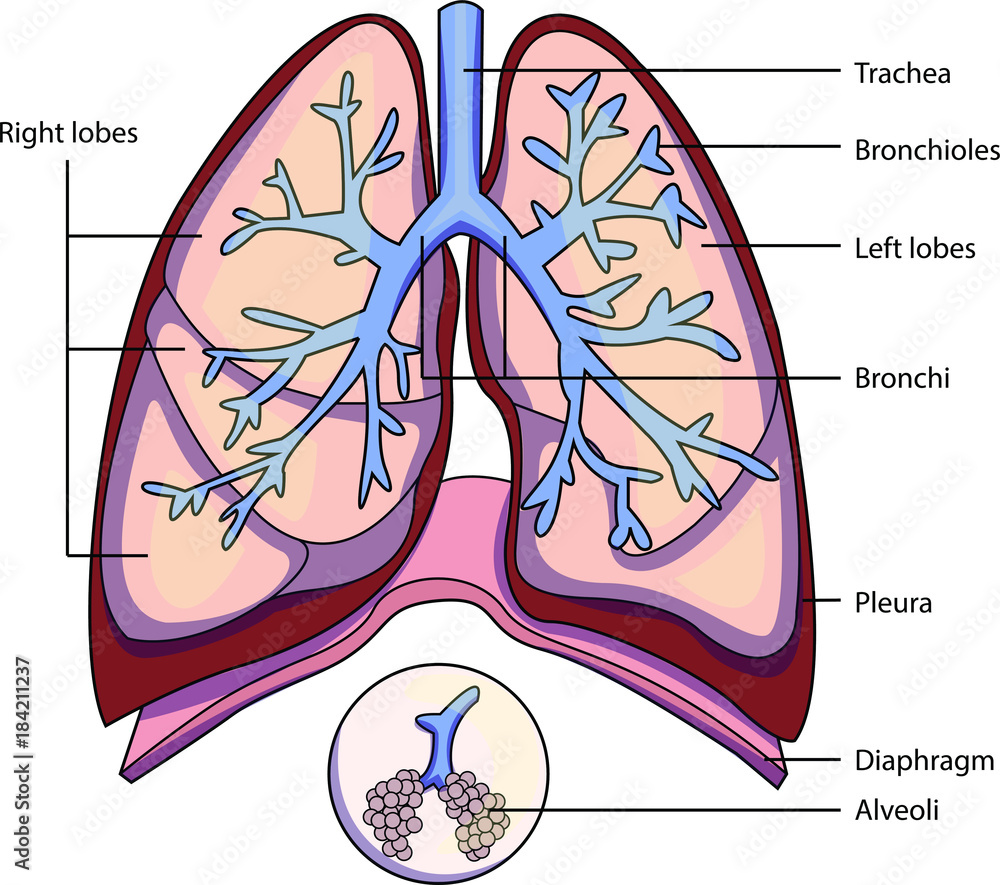
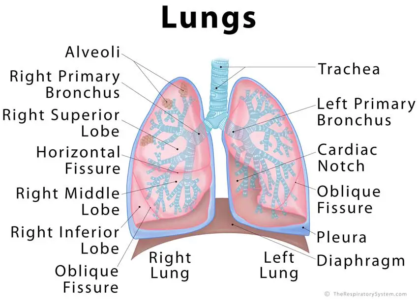
Diagram of the respiratory system. Air enters the body via the nose (preferably) or the mouth. The air enters the main windpipe, called the trachea, and continues en route to each lung via either the right or left bronchus (plural=bronchi). The lungs are separated into sections called lobes, two on the left and three on the right.
CrossFit Anatomy of the Lungs
Lung anatomy This spongy, pinkish organ looks like two upside-down cones in your chest. The right lung is made up of three lobes. The left lung has only two lobes to make room for your.
Images 07. Respiratory System and Breathing Basic Human Anatomy
Lung anatomy can get quite complicated extremely quickly. Ease into the topic and cement your knowledge using Kenhub's respiratory system quizzes and labeled diagrams. Regulation of breathing. The breathing cycle is controlled by the respiratory centre located inside the medulla oblongata and the pons of the brain stem. Three major collections.
Lung Anatomy & Function Lung Nodule, Lung Disease and Lung Infection
Lungs are a pair of respiratory organs situated in a thoracic cavity. Right and left lung are separated by the mediastinum. Texture -- Spongy Color - Young - brown Adults -- mottled black due to deposition of carbon particles Weight- Right lung - 600 gms Left lung - 550 gms THORACIC CAVITY SHAPE - Conical Apex (apex pulmonis) Base
Respiratory System Anatomy and Physiology Nurseslabs
the trachea the diaphragm The sections below will look at each part of the respiratory system in more detail. Nose and nasal cavity Forming the main external opening of the respiratory system,.
Lung Anatomy & Function Lung Nodule, Lung Disease and Lung Infection
Anatomy Structure There are two lungs (a right and left) in the body, but they are different sizes. The right lung is bigger and is divided into three lobes (separated by fissures), while the left lobe is smaller consisting of two lobes. The left lobe is also smaller as it has to make room for the heart.