
Turtle xray Stock Photo, Royalty Free Image 310764937 Alamy
view. Because sea turtles lack a diaphragm, the last two require horizontal placement of the x-ray beam to avoid ventral displacement of the lungs via the viscera. Make sure the patient is as near to the plate as possible to avoid magnification. Focal film distance (from machine to plate) should remain a consistent 40-42 inches.
Turtles Get New Digital X Ray System Jekyll Island Foundation
Reptile radiography is a useful diagnostic tool for various conditions in exotic pets. This pdf article provides an overview of the indications, techniques, and interpretation of reptile radiography, with examples of common findings and pitfalls.

Xray of a Turtle Shell Stock Image C028/0933 Science Photo Library
X-ray Microscopic Examination of a Turtle Figure 1: Maverick 3D rendering of volumetric data of turtle skeleton imaged with the SkyScan 1273 Micro-CT technology has transformed how zoologists conduct integrative and comparative biology research.

Turtle xray a photo on Flickriver
X-ray images of walking turtle in lateral (A) and ventral (B) views. Blue dots indicate markers on the pelvis, red dots are marker locations on the femur, and green dots are markers located on the.

xray of female turtle carrying eggs Vet Medicine, Veterinary Medicine, Freshwater Turtles
In addition, horizontal beam radiographs of turtles and tortoises are important for visualization of the lungs. Turtles can undergo x-ray exams awake, but general anesthesia is sometimes required to ensure good-quality positioning. Conscious turtles and tortoises have the ability to hold their extremities and head within or close to their shells.

TURTLE
Micro Photonics 204 subscribers Subscribe No views 1 minute ago Volumetric renderings from Bruker CTVox and Maverick Indie highlighting the skeletal structures of a turtle imaged using the.

Sea turtle xray Sea turtle, Turtle, Animals
Exotic Medicine Radiology/Imaging Reptile Radiography Issue: November/December 2014 Danielle Mauragis CVT Clifford R. Berry DVM, DACVR Radiography of reptile patients is routinely used for evaluation of traumatic injuries and the gastrointestinal and reproductive tracts. A reptile radiography study typically includes lateral and dorsoventral views.

Odd, eerie and cool Minnesota Zoo shares animal Xrays MPR News
Projections Due to the nature of the coelomic cavity, use horizontal-beam or cross-table projec-tions, if possible, for the lateral and cranio-caudal projections of turtles and tortoises. In other reptile species, vertical-beam dor-soventral projections are also acceptable. RADIOGRAPHIC EXPOSURE

Eastern Box Turtle Kingsbrook Animal Hospital's Blog
Show more Fluoroscopy of a turtle using 35 KV x-rays.NOTE: Nothing x-rayed that is alive is harmed during the process. The subject is placed into a custom container.

XRays Reveal A Surprise In This Turtle's Tummy Popular Science
No testudines (turtles and tortoises) are legally considered dangerous, but several species (eg,. A horizontal x-ray beam provides the best lateral imaging in lizards, especially when evaluating the respiratory system. The positioning for this view involves rotating the x-ray tube 90° and placing the cassette vertically behind the lizard.

One of my turtle graves xrayed X ray, Xray images, Sea shells
Download scientific diagram | (A, B) X-ray images of turtles during locomotion. (A) Cryptodire (P. concinna) walking, lateral view. (B) Pleurodire (E. subglobossa ) swimming, ventral view. Blue.

Sea Turtle XRay This is a photo of an XRay on display at… Flickr
Radiographic Positioning and Technique for Reptiles. Read this imaging article by Jodi Nugent-Deal discussing proper positioning and techniques for turtles, tortoises, snakes, and lizards.
This turtles xray r/Radiology
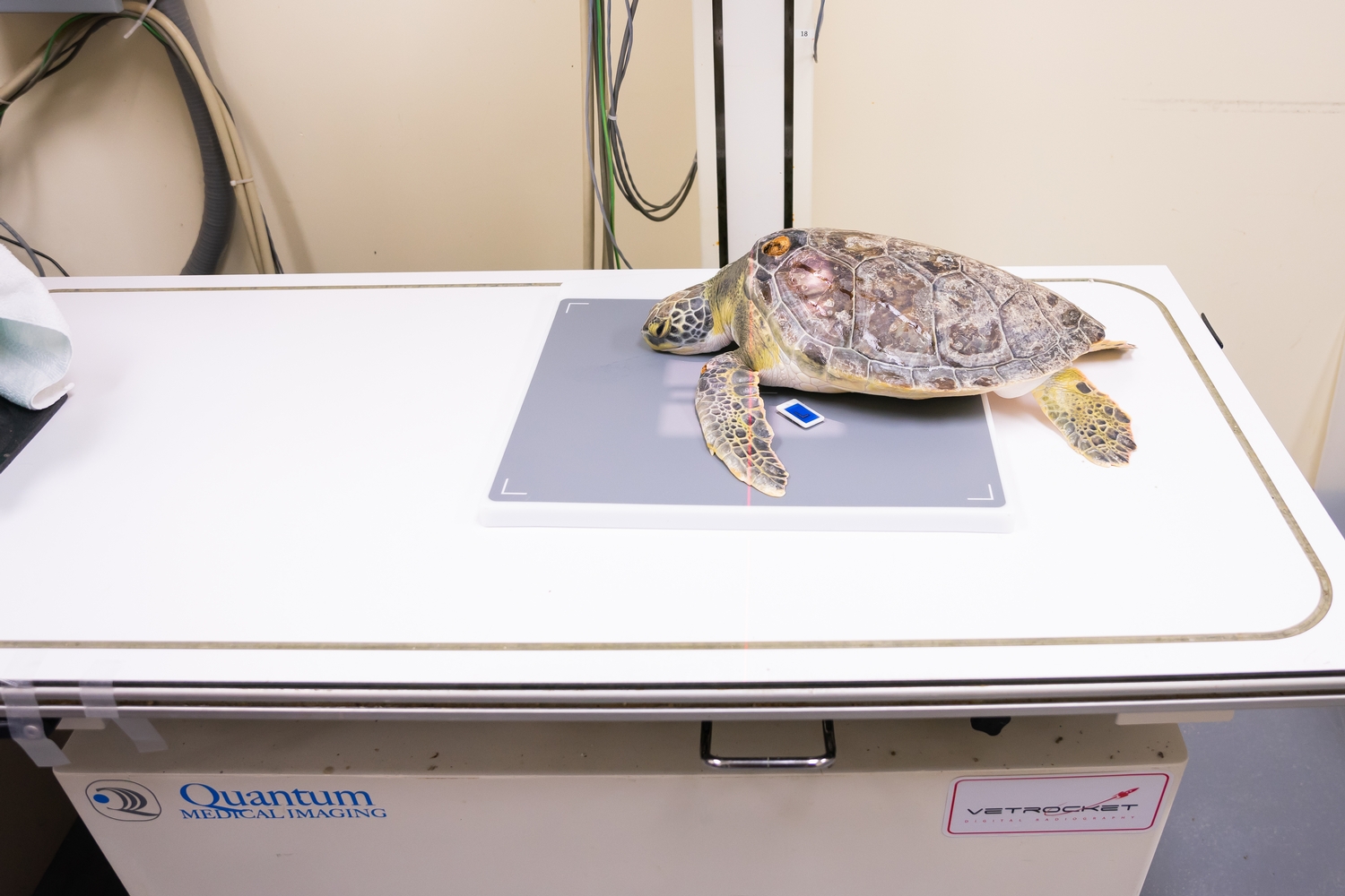
April 20, 2022 Turtle Radiography Here at the Wildlife Medical Clinic, we take digital radiographs, or x-rays, when we want to examine patients of all species and sizes. Radiographs are images of the body used to evaluate internal structures like organs and bones.
Turtle saved from painful fate by West Kelowna vet iNFOnews ThompsonOkanagan's News Source
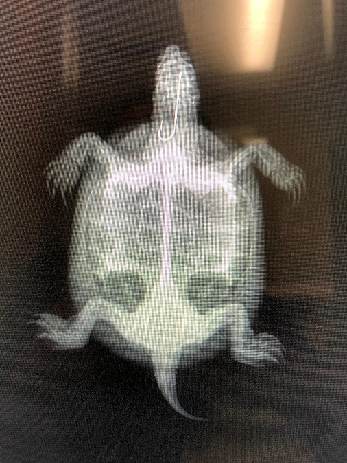
Xrays are needed for diagnosis of: Hook ingestion, location, and position Pneumonia Buoyancy disorders Pneumocoelom (free air in coelomic cavity) Trauma/fracture Osteomyelitis/arthritis Obstipation Prior to taking radiographs barnacles are removed from the carapace and plastron.

Fluoroscopic video of a turtle moving around, turtle xray, see insides of turtle HD 1080 YouTube
[ Read more ]. Ted Kinsman When Ted Kinsman picked up this specimen on the side of the road one spring morning, he was hoping to get a good X-ray image of a snapping turtle. But after he.
Fascinating Xray images of pregnant animals' bellies Daily Mail Online
Introduction. Reptilian species often show the same nonspecific symptoms with a variety of diseases. Radiologic studies in chelonians have been described by many authors (1-7).However, owing to the great variability of species, papers dealing with radiographic anatomy are limited to a representative number of species of each order of Reptilia ().In sea turtles, traumatic injuries of the.